
CT scan
A computerised tomography (CT) scan is a way to create detailed images of the inside of your body. It works by taking X-rays, which a computer processes to produce images. CT scans can diagnose and monitor various health conditions.
About CT scans
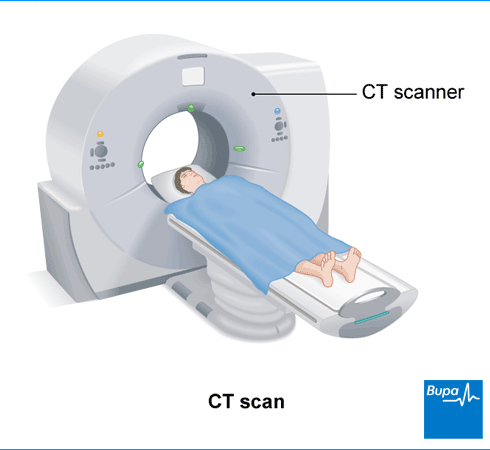
A CT scanner is a large, ring-shaped machine with a hole in its centre (like a doughnut shape). You lie on a table that moves through the middle of the machine. An X-ray tube inside the ring creates a beam of X-rays. As you lie flat on the table, the X-ray tube rotates around your body. The X-rays pass through your body and are picked up by detectors on the opposite side. These signals are then collected by a computer and built up into the detailed images of part of your body.
CT scans are usually taken across your body, which is called the axial plane. This is why a CT scan is sometimes referred to as a CAT scan (computed axial tomography scan). Scanners take the images continuously, like a spiral, in just a few seconds. CT scanners can also create 3D images.
Different parts of your body (such as bones and muscles) show up differently in the images from the CT scan. Bones will be white, whereas muscles may appear in different shades of grey. Sometimes the grey shades are very similar, which can make it difficult to tell different areas apart. A dye, called a contrast medium, can make the images clearer. You may have this as:
- an injection
- a liquid that you drink
- an enema (inserted into your bottom)
A radiographer – a health professional trained in performing CT and other scans – will operate the CT scanner. You usually have a CT scan as an out-patient in a hospital department. This means you have the scan and go home the same day.
Preparation for a CT scan
Before your appointment
Depending on the type of scan you’re having, your hospital may ask you not to eat or drink anything for several hours beforehand. If you’re having a CT scan of your large bowel, they may give you instructions on how to empty your bowel too. You might need to take laxatives, or follow a special diet for a couple of days. It’s important to follow any instructions your hospital gives you before your scan.
At the hospital
Your radiographer may ask you to change into a hospital gown for your CT scan. They may also ask you to remove any jewellery, glasses, contact lenses, dentures, hair clips and hearing aids. This is because metal can affect the images created by the scanner.
Your radiographer will discuss with you what will happen before, during, and after a CT scan. If you’re unsure about anything, don’t be afraid to ask. It’s important that you understand everything before you go ahead with the CT scan. Let your radiographer know if there’s a chance you might be pregnant.
Having contrast medium
If you need to have a CT scan with contrast dye, your radiographer will check whether you have any medical conditions, such as allergies, diabetes, or kidney disease. These could affect how you react to the contrast medium. If you have any problems with your kidneys, you may need to have some blood tests before your scan. Your radiographer may ask you to drink some water before your scan.
If you’re having a CT scan of your tummy (abdomen), your hospital will ask you to drink the contrast medium some time before your scan. You may need to drink some water before your scan too. You might need to come into hospital an hour and a half before your scan to do this.
CT scan procedure
Your radiographer will ask you to lie on the CT scanner table, usually flat on your back. They’ll provide any necessary supports or pillows to help you stay in position comfortably and keep still.
If you’re having contrast medium, your radiographer will inject this through a small tube (cannula) into your arm. It may make you feel warm for a minute or two, and it can cause a metallic taste in your mouth. You may also feel like you need to pee but this wears off quickly.
Your radiographer will control the position of the table, and will move it into the right place to start the scan. The table will then move through the machine as your radiographer takes the scan. The CT scan itself usually only takes a few minutes. It’s important to lie very still and you may need to hold your breath for a short time – your radiographer will guide you through this. You may hear some whirring noises from the CT scanner when you’re inside it.
Your radiographer will operate the scanner from a control room behind a window but they’ll be able to see, hear and speak to you throughout. A CT scan isn’t painful, but you might feel a bit uncomfortable from having to stay still for a few minutes.
Uses of a CT scan
Doctors often prefer CT scans to standard X-rays, because they give clearer, more detailed images. They can be used to detect or monitor many health conditions including:
- stroke
- broken bones or other problems that affect your bones
- cancers
- infection, inflammation and other damage inside your body
- conditions that affect your heart and blood vessels – this type of CT scan is called a CT angiography or CT venography
- digestive problems that affect your stomach and intestines – a CT scan of your large bowel is called a virtual colonoscopy
- problems that affect your kidneys, urinary system or bladder – this is called a CT pyelography or CT urography
Doctors may also use CT scans when planning surgery, or to guide them during procedures, such as biopsies.
Aftercare for CT scan
Once the scan is finished, your radiographer will come back into the room and lower the table so you can get down. If you had an intravenous (IV) contrast, they’ll remove the cannula from your arm. You’ll need to stay in the radiology department for 15 to 30 minutes if you’ve had IV contrast, to make sure you don’t have any side-effects. Otherwise, you’ll be able to go home as soon as you feel ready.
Before you go home, ask your radiographer when you can expect to get your results. It can take a week or 2 for them to come through.
A radiologist (a doctor who specialises in using imaging methods to diagnose medical conditions) will review your results. They’ll usually send a report to the doctor who referred you for the CT scan.
Complications of CT scan
As with every test, CT scans have some risks. But the benefits of having a CT scan usually outweigh these risks. Your radiographer will explain these to you.
Radiation
A CT scan, like any radiology test, exposes you to some radiation. Radiation can be harmful to your body if you have a high enough dose. The amount of radiation you get from a CT scan is more than that from other types of X-ray imaging, such as a standard X-ray. Your doctor and radiographer will keep your exposure to a minimum. They’ll only recommend a CT scan when the benefit of having the scan outweighs the risk. Sometimes, they may recommend an alternative test.
If you're pregnant, there’s a risk the radiation could harm your baby. How big a risk there is can depend on how many weeks pregnant you are, and the area of your body being scanned. Your doctor may still suggest you have a CT scan if it’s an emergency, or if they think the benefits would outweigh any potential risks.
Contrast medium
Your radiographer will use a dye (contrast medium) for some CT scans. If you’ve had the contrast medium by injection (intravenously), you may have some discomfort as you have the injection. It can also make you feel or be sick (vomit).
Although very rare, it’s possible to have an allergic reaction to the contrast medium. If you get an itchy rash or have any difficulty breathing, tell your radiographer straightaway. Any reaction will usually happen within 15 minutes of having the contrast medium. Radiology departments are well set up to deal with these reactions. You may be at greater risk of an allergic reaction if you have asthma.
If you have kidney disease or diabetes, the contrast dye can make your kidney condition worse. Your doctor will ask you about any health conditions you have before the scan, so that they can take measures to reduce your risk.
Alternatives to CT scan
A CT scan tends to give more complete pictures of your organs and tissues than an ultrasound or X-ray. It’s also quicker than a magnetic resonance imaging (MRI) scan and you can have one even if you have an implanted medical device. But sometimes, an X-ray, ultrasound or MRI may be a better option. This depends on your own individual circumstances, as well as the reason why you’re having the scan. Other types of scan are often a better option for babies and children, or if you’re pregnant.
Health checks for peace of mind
A Bupa health check can help rule things out, so you can stop asking “but what if?”. No insurance required.
CT scans can detect or monitor health conditions ranging from strokes and broken bones, to cancer and problems with your digestive system. Doctors may also use CT scans before surgery to gather information, or to guide them during procedures such as biopsies.
See our uses of a CT scan section for more information.
No, you won’t get your CT scan results straightaway. A specialist doctor will review the results and send a report to the doctor who referred you for a CT scan. This can take up to a week or two.
No, a CT scan isn’t painful although you may feel a bit uncomfortable staying still while the scan is happening.
See our CT scan procedure section for more information.
The CT scan usually takes only a few minutes, but you may need to stay around for a little longer after the scan. For example, if your radiographer gave you a contrast medium, you’ll need to wait to check you haven’t had any reaction to it.
See our aftercare for CT scan section for more information.
CT scans and MRI scans differ in how they work. A CT scan works by taking X-rays which a computer processes to produce images. An MRI scan uses a powerful magnet and radio waves to create images. A CT scan uses a large, ring-shaped machine with a hole in its centre (like a doughnut-shape). Whereas if you have an MRI scan, you’ll usually go into a tube-shaped machine.
Related information
MRI scan
An MRI scan is a type of test you can have to produce detailed images of the inside of your body
Ultrasound
Ultrasound is a type of scan that uses sound waves to produce images of the inside of your body
X-rays
An X-ray machine works by projecting a beam of X-rays through the part of your body that your doctor needs to look at
Other helpful websites
Discover other helpful health information websites.
Did our CT scan information help you?
We’d love to hear what you think. Our short survey takes just a few minutes to complete and helps us to keep improving our health information.
The health information on this page is intended for informational purposes only. We do not endorse any commercial products, or include Bupa's fees for treatments and/or services. For more information about prices visit: www.bupa.co.uk/health/payg
This information was published by Bupa's Health Content Team and is based on reputable sources of medical evidence. It has been reviewed by appropriate medical or clinical professionals and deemed accurate on the date of review. Photos are only for illustrative purposes and do not reflect every presentation of a condition.
Any information about a treatment or procedure is generic, and does not necessarily describe that treatment or procedure as delivered by Bupa or its associated providers.
The information contained on this page and in any third party websites referred to on this page is not intended nor implied to be a substitute for professional medical advice nor is it intended to be for medical diagnosis or treatment. Third party websites are not owned or controlled by Bupa and any individual may be able to access and post messages on them. Bupa is not responsible for the content or availability of these third party websites. We do not accept advertising on this page.
- Patel PR, De Jesus O. CT scan. StatPearls Publishing.. www.ncbi.nlm.nih.gov/books, last updated 2 January 2023
- Computed tomography. MSD Manual Professional Version. www.msdmanuals.com, reviewed/revised April 2021
- Computerised tomography (CT) scans. Patient. patient.info, last edited 19 October 2021
- Conventional radiography. MSD Manual Professional Version. www.msdmanuals.com, reviewed/revised April 2021
- CT scan. Cancer Research UK. www.cancerresearchuk.org, last reviewed 5 August 2022
- Your CT scan. Royal College of Radiologists. www.rcr.ac.uk, accessed 28 August 2023
- Radiographic contrast agents and contrast reactions. MSD Manual Professional Version. www.msdmanuals.com, reviewed/revised April 2021
- Personal communication, Dr Paul Crowe, Consultant Interventional Radiologist, 22 September 2023
- Van Dam LF, Van Walderveen MAA, Kroft LJM, et al. Current imaging modalities for diagnosing cerebral vein thrombosis – a critical review. Thromb Res 2020; 189:132–39. doi: 10.1016/j.thromres.2020.03.011
- What does a clinical radiologist do? Royal College of Radiologists. www.rcr.ac.uk, accessed 28 August 2023
- Risks of medical radiation. MSD Manual Professional Version. www.msdmanuals.com, reviewed/revised April 2021
- Anaphylaxis. MSD Manual Professional Version. www.msdmanuals.com, reviewed/revised October 2022
- Magnetic resonance imaging. MSD Manual Professional Version. www.msdmanuals.com, reviewed/revised April 2021
- MRI scan. Macmillan. www.macmillan.org.uk, reviewed 27 January 2022