
MRI scan
MRI stands for magnetic resonance imaging. An MRI scan is a type of test that creates detailed images of the inside of your body. It uses a powerful magnet and radio waves and is often used to diagnose or monitor a condition.
What is an MRI scan used for?
An MRI scan can be used to look at almost any part of your body. It can detect many problems such as inflammation, tumours, or a torn ligament. The pictures created by an MRI scan can help to show any differences between healthy and unhealthy tissue.
An MRI scan can be used to diagnose or monitor:
- conditions that affect your brain, spine, and nervous system, such as brain tumours, spinal disc problems, and multiple sclerosis
- heart conditions such as valve problems
- problems with your blood vessels, such as narrowing of your arteries or risk of rupture (bursting)
- cancer
- liver or kidney disease
- conditions or injuries that affect your joints, ligaments or muscles such as arthritis or sports injuries
- bowel conditions such as Crohn’s disease or ulcerative colitis
How does an MRI scan work?
Images are taken in thin ‘slices’ through your body from various directions without you having to move. An MRI scan can often show things that can’t be seen on an X-ray or an ultrasound scan.
Some MRI scans use a special dye called a contrast medium to create more detailed pictures. The dye is injected into your body, usually through a vein in your arm.
Who can have an MRI scan?
An MRI scan has lots of benefits, but it isn’t suitable for everyone. The magnetic field from an MRI scanner can affect some metals, including those used in surgical clips or pins. If you’ve had surgery that left metals inside your body, it’s important to tell your radiographer (a healthcare professional who specialises in imaging). This is because the magnets can make the metal move, which could damage nearby tissues.
The magnetic field from the MRI may also affect electronic implants such as pacemakers. This may mean the devices don’t work properly or may heat up, which could present a risk. You can’t usually have an MRI scan if you have:
- a heart pacemaker or defibrillator (a device that keeps your heart rhythm regular)
- an inner ear hearing aid (cochlear implant)
- an aneurysm clip (a metal clip on an artery in your brain)
If you’re pregnant, you won’t usually be offered an MRI scan, especially during the first three months, but sometimes they are used to see if your baby has any congenital heart conditions (heart conditions present from birth). Doctors don’t know for sure if the magnetic field of an MRI could affect your unborn baby. But if you’re pregnant or think you may be, tell your radiographer before your MRI appointment. If you do need to have an MRI scan while you’re pregnant, the scan will be done in a way that reduces any risk to your baby.
If you’re claustrophobic, tell your doctor. You may find it difficult to have an MRI scan because the machine that you lay in is shaped like a tunnel. But there are things that can help, such as taking deep breaths, listening to music, or in some cases, taking a sedative. In some locations, open scanners are available for patients whose claustrophobia cannot be managed.
Preparing for an MRI scan
Before the day of your MRI scan
Before having an MRI scan, you should be able to eat, drink and take any medicines as usual. But your radiographer will give you specific instructions.
If you wear stick-on medicine patches, you may need to take these off before having an MRI scan, so take a spare one to the hospital. This is because some patches contain metal and may heat up during the scan.
On the day of your MRI scan
At the hospital, your radiographer will ask you to complete and sign a safety questionnaire. You’ll be asked if you’re pregnant or have any medical conditions. You’ll also be asked if you have any metal or electronic implants.
Your radiographer will go through a checklist of metal items that you may have inside your body. Having something metallic in your body doesn’t necessarily mean you can’t have an MRI scan. But your radiographer does need to know, so they can make the right safety decisions.
Before you go into the scanning room, you’ll probably be asked to undress down to your underwear and put on a hospital gown. You’ll probably be asked to remove your:
- underwired bra (which usually contains metal)
- jewellery (including piercings)
- watch
- metal hairclips
- dentures with metal parts
- glasses and hearing aids if you wear them
- make-up, especially eye make-up, which can contain metal fibres
Tell the radiographer if you have any tattoos, especially if they’re on the part of your body that’s being scanned. Your tattoo may need to be covered with a cold compress or ice pack during the scan. This is because tattoos can heat up if they contain any metal.
Don't take any electronic or metal items, such as your keys, mobile phone or credit cards, with you into the scanning room. A friend or relative may be able to stay with you during the scan. They’ll also have to leave any metal or electronic items behind, and complete and sign a safety questionnaire.
Young children and people with claustrophobia may be offered a sedative or a general anaesthetic before an MRI scan. if they’re doctor thinks this will help them to lie still.
Before you have an MRI scan with a dye
If you’re having an MRI scan that uses a dye (contrast medium), your doctor may need to inject this before or during your scan. Where it’s injected will depend on which part of your body needs scanning and why. You may need a blood test before your scan to check the dye is right for you.
You may not be able to have the dye if your kidneys aren’t working well. This may be because you:
- have diabetes or heart failure
- are very dehydrated
- are taking medicines that affect your kidneys
A very small number of people may be allergic to the dye. Tell your radiographer before your scan if you have any allergies.
What are the alternatives to an MRI scan?
If an MRI scan isn’t suitable for you, you may be able to have other tests that create images of the inside of your body. These include:
- X-ray – this uses radiation to create an image
- ultrasound – this uses sound waves to create an image
- computerised tomography (CT) scan – this uses X-ray equipment and computer software to create images
Your doctor will discuss your options and recommend the best test for you.
What happens during an MRI scan?
You’ll usually have your MRI scan and go home on the same day.
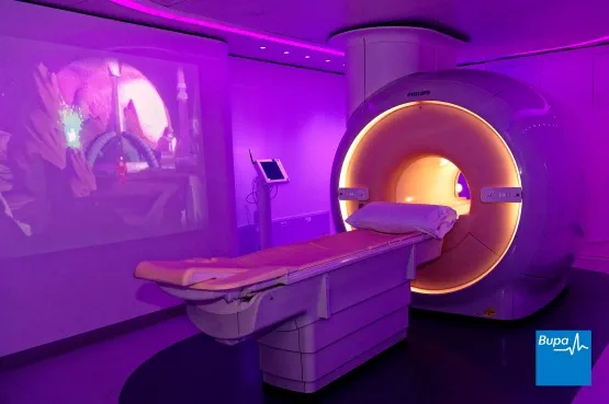
When you go for your MRI scan, your radiographer will ask you to lay on the scanner table. The table slides into the cylinder-shaped MRI scanner, which contains a magnet. Your position may be adjusted using foam pads. Your radiographer will make sure you’re comfortable and any equipment around you is in a safe place.
You’ll be told before your scan how long it’s likely to take. You’ll usually need to lie still for about 20 minutes, sometimes with your hands at your sides. Your scan may take longer than this depending on how much of your body is being scanned. You’ll be encouraged to close your eyes and relax as much as possible.
Making the MRI scan more comfortable
Your radiographer will be in a separate room during the scan, but there is a window between the control room and the room you are in. They may ask you to hold your breath at certain times during the scan. They’ll be able to see and hear you during the scan, and you can talk to them if you need to. An MRI scanner is very noisy and makes knocking or drilling sounds1. This is the sound of the magnetic fields changing during the scan.
To make an MRI scan more comfortable for you, your radiographer will:
- talk to you regularly to reassure you and remind you to be as still as possible
- show you how to use the buzzer if you need to talk to them
- give you earplugs or headphones to wear or may play background music for you to listen to if you wish
- discuss with you what can be done to reduce any anxiety, especially if you have claustrophobia
An MRI scan can sometimes increase your body temperature or make you feel warm. If you feel burning or discomfort in any part of your body during the test, tell your radiographer straight away.
What to expect afterwards
After your MRI scan, you’ll usually be able to go home as soon as you feel ready. You should be able to go back to your normal routine straightaway.
A radiologist (a doctor who specialises in using imaging methods to diagnose medical conditions) will examine your MRI scans. The results will then be sent to the doctor who requested the test. You’ll have a follow-up appointment to find out the results.
Looking for prompt access to quality care?
With our health insurance, if you develop new conditions in the future, you could get the help you need as quickly as possible, from treatment through to aftercare.
To get a quote or to make an enquiry, call us on 0800 600 500∧
Having tooth implants or fillings shouldn’t stop you from having an MRI scan. But it’s important to let your radiographer know if you have any of these so they can make the right decision. Metal tooth implants and crowns may affect the pictures created by the MRI scanner if you’re having your head scanned – you can check with your dentist to find out if there is metal in your teeth.
There are lots of differences between MRI scans and CT scans. An MRI scan takes images of your soft tissues and doesn’t use any radiation. CT scans use radiation and can sometimes be a quicker procedure. Your doctor will be able to decide which is the appropriate option for your needs.
If you’re claustrophobic, you may still be able to have an MRI scan. Although MRI scanners resemble a tunnel, they are open at either end so you won’t be completely enclosed at any time. You can communicate with your radiographer via a buzzer if you feel uncomfortable during the procedure. And they may recommend you listen to music or use a sedative if necessary. Contact the radiography department where you’re having your scan when your appointment comes through to discuss your options.
CT scan
A CT scanner is a large, ring-shaped machine used to diagnose and monitor a number of different health conditions
Ultrasound
Ultrasound is a type of scan that uses sound waves to produce images of the inside of your body
X-rays
An X-ray machine works by projecting a beam of X-rays through the part of your body that your doctor needs to look at
General anaesthesia
General anaesthesia is when medication is given to make you temporarily unconscious during an operation, so don't feel pain or other sensations.
Other helpful websites
Discover other helpful health information websites.
Did our MRI scan information help you?
We’d love to hear what you think. Our short survey takes just a few minutes to complete and helps us to keep improving our health information.
The health information on this page is intended for informational purposes only. We do not endorse any commercial products, or include Bupa's fees for treatments and/or services. For more information about prices visit: www.bupa.co.uk/health/payg
This information was published by Bupa's Health Content Team and is based on reputable sources of medical evidence. It has been reviewed by appropriate medical or clinical professionals and deemed accurate on the date of review. Photos are only for illustrative purposes and do not reflect every presentation of a condition.
Any information about a treatment or procedure is generic, and does not necessarily describe that treatment or procedure as delivered by Bupa or its associated providers.
The information contained on this page and in any third party websites referred to on this page is not intended nor implied to be a substitute for professional medical advice nor is it intended to be for medical diagnosis or treatment. Third party websites are not owned or controlled by Bupa and any individual may be able to access and post messages on them. Bupa is not responsible for the content or availability of these third party websites. We do not accept advertising on this page.
- Magnetic resonance imaging. Patient. patient.info, last reviewed January 2018
- Magnetic resonance imaging. The MSD Manuals. www.msdmanuals.com, last full review/revision September 2025
- Ischemic stroke. The MSD Manuals. www.msdmanuals.com, last updated August 2023
- Multiple sclerosis (MS). The MSD Manuals. www.msdmanuals.com, last full review/revision October 2025
- Musculoskeletal conditions. Oxford Handbook of Adult Nursing. Oxford Medicine Online. oxfordmedicine.com, published online June 2018
- Colorectal assessment. OSH Colorectal Surgery. Oxford Medicine Online. oxfordmedicine.com, published online October 2011
- Safety guidelines for magnetic resonance imaging equipment in clinical use. MHRA. www.gov.uk, updated April 2022
- Brain magnetic resonance scanning. Medscape. emedicine.medscape.com, updated November 2020
- MRI scans. British Health Foundation. www.bhf.org.uk, published September 2023
- Sedation explained. Royal College of Anaesthetists. 2018. www.rcoa.ac.uk, accessed February 2020
- Plain abdominal X-ray. Patient. patient.info, last reviewed February 2016
- Ultrasound scanning – non-obstetric. Patient. patient.info, last reviewed December 2015
- Computerised tomography (CT) scans. Patient. patient.info, last reviewed July 2015
- Chockattu SJ, Deepak BS, Thakur S. Unwanted effects due to interactions between dental materials and magnetic resonance imaging: a review of the literature. Restor Dent Endod 2018; 43(4):e39. doi:10.5395/rde.2018.43.e39
- Görgülü S, Ayyildiz S, Kamburoğlu K, et al. Effect of orthodontic brackets and different wires on radiofrequency heating and magnetic field interactions during 3-T MRI. Dentomaxillofac Rad 2014; 43(2):20130356. doi:10.1259/dmfr.20130356
- Mathew CA, Maller S, Maheshwaran M. Interactions between magnetic resonance imaging and dental material. J Pharm Bioallied Sci 2013; 5(Suppl 1):S113–16. doi: 10.4103/0975-7406.113309
- Personal communication, Julia Ross, Radiographer and Head of MSK and Radiology at Bupa, September 2020