
Ultrasound
An ultrasound is a type of scan that uses sound waves to create images of the inside of your body. It can pick up changes in your organs, tissues, blood vessels, and joints. This may help to diagnose some health problems. Ultrasound scans are also used in certain procedures and during pregnancy.
About ultrasound
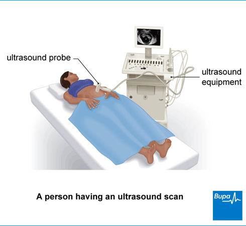
An ultrasound machine consists of a small hand-held scanner (‘probe’). The probe contains a transducer that sends out sound waves. The sound waves are a high frequency that you can’t hear. They bounce off the organs and tissues inside your body and are picked up again by the probe. A layer of water-based gel is placed on your skin under the probe. This helps with the transmission of the sound waves.
The probe is linked to a computer that creates real-time images from the reflected sound waves. The images are displayed on a monitor in various shades of grey. The pictures are constantly updated. This means the scan can show movement. The pictures can be captured, saved, and shared with you.
Hand-held ultrasound machines are much smaller and easier to transport than the computer-based versions. Several different types of healthcare professional may carry out ultrasound scans. These include sonographers (specialists in ultrasound) and radiologists (doctors specialising in imaging methods).
Ultrasound uses
You can have an ultrasound on different parts of your body. These include your:
- muscles and joints – to check for an injury or health conditions such as osteoarthritis
- tummy (abdomen) – to check the health of organs such as your liver, gallbladder, kidneys, and pancreas
- reproductive organs such as your womb and ovaries, or prostate and testes
- breasts – to check lumps, alongside other tests such as mammograms and biopsies
- lumps under your skin, anywhere around your body
- blood vessels – to check for conditions such as deep vein thrombosis, varicose veins, or abdominal aortic aneurysm, or to assess your risk of stroke
You may also have an ultrasound to:
- help diagnose cancer
- check the health of your baby when you’re pregnant
- monitor infertility treatments
Ultrasound is also used to help doctors and surgeons perform certain procedures. It helps your doctor to guide the needle to the right place. These procedures include:
- biopsy, which involves taking a sample of cells using a small needle
- steroid joint injections
The healthcare professional referring you will explain why they’re recommending you have an ultrasound scan. If you have any questions, make sure you ask them. You’ll need to give your consent when you go for the scan.
Types of ultrasound
There are three main types of ultrasounds: external, internal, and endoscopic.
External ultrasound
The ultrasound practitioner moves the ultrasound probe over your skin.
Internal ultrasound
The ultrasound practitioner places the probe inside your body to take the scan. This may be:
- a transvaginal ultrasound in your vagina to check your womb and ovaries
- a transrectal ultrasound in your back passage to check your prostate, if you have one, or to take images inside your back passage
Endoscopic ultrasound
This is a combination of an endoscopy (gastroscopy) and an ultrasound scan. The practitioner uses a probe attached to a long, thin, flexible tube (an endoscope) to look further inside your body. The probe is passed into your mouth and down your food pipe (oesophagus) to check for problems in your digestive system.
Special types of ultrasounds
Some types of ultrasounds use special techniques. These include the following.
- Echocardiogram. This is an ultrasound of your heart. An echocardiogram can create real-time images of blood flowing through your heart.
- Doppler (or Duplex) ultrasound. This can monitor the blood flow in your blood vessels. It can show how fast and in which direction your blood is flowing. This can help to detect blood clots or narrowed blood vessels.
- 3D ultrasound. Standard ultrasound creates 2D (flat) images. But newer technology is also able to create 3D ultrasound images. This may make it easier to see any problems.
- Elastography. This can measure the stiffness of organs inside your body. It’s most commonly used to check if you have liver fibrosis (scarring). Fibrosis can be due to alcohol-related liver disease, non-alcoholic fatty liver disease, and hepatitis.
- Contrast-enhanced ultrasound. This uses contrast agent (dye) to show up your vessels or organs more clearly. Your doctor or sonographer injects very small gas bubbles with the contrast agent into your vein before the procedure.
Preparation for ultrasound
Whether or not you need to do anything to prepare for your ultrasound depends on the type of ultrasound you’re going to have. For some scans, you may need to remove some clothing. So, it’s usually helpful to wear loose-fitting clothes that you can take off easily.
For some types of ultrasounds, you may be asked to follow specific instructions about eating or drinking before your procedure. This is to make it easier for the practitioner to create clear images.
If you’re having a scan of your womb, bladder, or kidneys, you may need to drink something before your appointment, so you have a full bladder. Sometimes you may then be asked to go for a pee, so the ultrasound can be repeated when you have an empty bladder.
If you’re having a scan of your gallbladder or pancreas, you may be asked to stop eating for about six hours before the scan.
If you’re having a transrectal ultrasound, you may be asked to make sure your bowel is empty before your appointment.
If you’re having an endoscopic ultrasound, you’ll usually need to stop eating at least six hours beforehand, so your bowel is clear. You can usually carry on drinking for two hours before the test. You may need to have more preparation as part of having a gastroscopy.
Ultrasound procedure
An ultrasound appointment can take anything from 10 minutes to 30 minutes. This will depend on why you’re having it. The person doing your scan will explain the procedure to you and check you’re happy to go ahead. You may be asked to remove clothing or change into a hospital gown. This depends on which part of your body is being scanned.
If you’re having an abdominal ultrasound, you’ll usually need to lie on your back on a couch. Your ultrasound practitioner will put some gel on your skin on the area they’re going to scan. This may feel cold. The gel helps to transmit the soundwaves between your body and the probe.
The practitioner will hold the probe firmly against your skin and move it over the surface. They may ask you to take some deep breaths or to move into different positions so they can get the best possible images.
An ultrasound isn’t painful, but you may feel some slight discomfort when the probe is pressed against you, particularly if the area is tender. Tell the person doing your scan if you feel uncomfortable.
After an ultrasound, your practitioner will usually wipe the gel off your skin. If any is left, it won’t stain your clothing. You’ll be able to wash it off with soap and water when you get home.
Transvaginal ultrasound
If you’re having a transvaginal ultrasound, the ultrasound practitioner will ask you to lie on your back with your knees raised and legs apart. If this is difficult for you, you may be able to lie on your side with your knees pulled up to your chest instead.
The practitioner inserts a small, thin ultrasound probe (similar in size to a tampon) into your vagina. They’ll put a protective cover over the probe. It will have some gel on it. It’s important to tell your hospital if you have a latex allergy so they can use a suitable cover.
The test may be a bit uncomfortable, but it shouldn’t be painful.
Transrectal ultrasound
If you’re having a transrectal ultrasound, your practitioner will ask you to lie on your left side with your knees pulled up towards your chest. Your practitioner will insert a small, thin ultrasound probe into your back passage. The probe will have a protective cover and lubricating gel on it. If you have a latex allergy, tell the practitioner so they can use a suitable cover.
A transrectal ultrasound may feel a bit uncomfortable but it shouldn't be painful.
Endoscopic ultrasound
You’ll usually have a sedative for an endoscopic ultrasound. This will make you feel relaxed and sleepy, but you’ll still be awake.
The healthcare professional performing the test may spray some local anaesthetic on your throat to numb it. They’ll then pass the endoscope through your mouth and guide it towards the area of your body that’s being scanned. You may need to swallow as the tube goes down. Once the practitioner has taken the images they need, they’ll gently remove the endoscope.
Aftercare for ultrasound
After an ultrasound, you can usually go straight home or continue with your usual activities. If you’ve had a sedative, you may need to rest for a while until the effects have worn off. Ask a friend or family member to take you home by car or taxi – ideally not public transport. The effects of sedation can last up to 24 hours, so ask them to stay overnight to look after you. Don’t drive, drink alcohol, operate machinery, or make any important decisions during this time. Your hospital will give you more information about having a sedative before your scan.
During an endoscopic ultrasound, your doctor may have used a spray to numb your throat. It’s best to wait about an hour for this to wear off before you eat or drink anything. You might have a sore throat for a few days afterwards.
Getting your ultrasound results
Ultrasound scan results will usually be sent to the healthcare professional who referred you for the scan. They will then discuss the results with you. Ask them how long it should take to get the results.
Risks of ultrasound
An ultrasound is considered safe because it doesn’t use any radiation. So there aren’t any of the risks associated with scans such as X-rays and CT scans, which do use radiation. Even so, ultrasound scans should only be done for clear medical reasons.
Alternatives to ultrasound
Alternatives to ultrasound imaging depend on why you’re having the scan. It will also depend on which part of your body is being looked at and your individual circumstances. Alternative scans may include:
You may have ultrasound as well as these other scans. Your doctor will talk you through which type of scan is most suitable for you.
GP Subscriptions – Access a GP whenever you need one for less than £20 per month
You can’t predict when you might want to see a GP, but you can be ready for when you do. Our GP subscriptions are available to anyone over 18 and give you peace of mind, with 15-minute appointments when it suits you at no extra cost.
You may have an ultrasound to help diagnose a health condition or injury. The scan can pick up changes in your organs, tissues, blood vessels, or joints. You may also have an ultrasound during a medical procedure, or to check your baby during pregnancy.
See our Ultrasound uses section for more information.
You don’t always need to prepare for an ultrasound scan, but it’s a good idea to wear loose clothing. Sometimes, you may be asked to follow specific instructions. You may need a full bladder for your scan. Or you may need to stop eating and drinking for a certain number of hours beforehand. See our Preparation for ultrasound section or more information.
See our Preparation for ultrasound section for more information.
If you’re having a pregnancy ultrasound scan, the healthcare professional carrying out your scan may be able to discuss the results straight away. But other types of ultrasound scan results will usually be sent to the doctor or healthcare professional who referred you for the test.
For more information, see our Aftercare for ultrasound section.
Did our Ultrasound information help you?
We’d love to hear what you think. Our short survey takes just a few minutes to complete and helps us to keep improving our health information.
The health information on this page is intended for informational purposes only. We do not endorse any commercial products, or include Bupa's fees for treatments and/or services. For more information about prices visit: www.bupa.co.uk/health/payg
This information was published by Bupa's Health Content Team and is based on reputable sources of medical evidence. It has been reviewed by appropriate medical or clinical professionals and deemed accurate on the date of review. Photos are only for illustrative purposes and do not reflect every presentation of a condition.
Any information about a treatment or procedure is generic, and does not necessarily describe that treatment or procedure as delivered by Bupa or its associated providers.
The information contained on this page and in any third party websites referred to on this page is not intended nor implied to be a substitute for professional medical advice nor is it intended to be for medical diagnosis or treatment. Third party websites are not owned or controlled by Bupa and any individual may be able to access and post messages on them. Bupa is not responsible for the content or availability of these third party websites. We do not accept advertising on this page.
- Ultrasonography. The MSD Manuals. www.msdmanuals.com, modified March 2024
- Non-obstetric ultrasound scanning. Patient. patient.info, last updated December 2021
- Going for an ultrasound scan. British Medical Ultrasound Society. www.bmus.org, accessed July 2025
- Guidelines for professional ultrasound practice. 8th edition. Society of Radiographers and British Medical Ultrasound Society. www.bmus.org, published December 2023
- Ultrasound scan. Cancer Research UK. www.cancerresearchuk.org, last reviewed November 2022
- Colorectal cancer. BMJ Best Practice. bestpractice.bmj.com, last reviewed June 2025
- What to expect from different types of ultrasound examination. British Medical Ultrasound Society. www.bmus.org, accessed July 2025
- Echocardiography. The MSD Manuals. www.msdmanuals.com, reviewed/revised December 2023
- Upper and lower limb venous duplex ultrasound examination for the assessment of deep vein thrombosis (DVT). The Society for Vascular Technology of Great Britain and Ireland. www.csvs.org.uk, reviewed March 2021
- Iommi D, Hummel J, Figl ML. Evaluation of 3D ultrasound for image guidance. PLoS One. 2020 Mar 26;15(3):e0229441. doi: 10.1371/journal.pone.0229441. PMID: 32214326; PMCID: PMC7098612
- Scans and imaging tests. British Liver Trust. britishlivertrust.org.uk, accessed July 2025
- Guo S, Gan L, Du F, et al. S index: a predictor comparable to ultrasound shear wave elastography in assessing liver fibrosis in patients with chronic hepatitis B. BMC Med Imaging 25, 296 (2025).
- Cirrhosis. Patient. patient.info, last updated November 2024
- Kurzweil A, Martin J. Transabdominal ultrasound. StatPearls Publishing. www.ncbi.nlm.nih.gov, last update August 2023
- Endoscopic ultrasound. Cancer Research UK. www.cancerresearchuk.org, last reviewed June 2025
- Sedation explained. 3rd edition. Royal College of Anaesthetists. rcoa.ac.uk, published March 2025
- Caring for someone recovering from general anaesthetic or sedation. 3rd edition. Royal College of Anaesthetists. rcoa.ac.uk, published March 2025
- Gastroscopy. Cancer Research UK. www.cancerresearchuk.org, last reviewed June 2025
- Diagnostic imaging and scans. Patient. patient.info, last updated August 2023
